STRONG SCIENCE

STATE OF THE ART TECH

The technology behind Mpower has been designed with one thing in mind: make people stronger. To ensure we can stay 100% behind our words, we have build everything based on science. So, if you are interested in knowing more, this is for you.


1. WHAT IS sEMG SIGNAL?


EMG (electromyography) consists of electrical signals of nerves and muscles controlling the contraction of the muscles

• EMG can be measured non-invasively with special electrodes placed on the skin overlying the muscle(s) of interest (i.e. surface EMG, sEMG)

• sEMG technology has enabled the use of sEMG signal for medical and sports physiology related muscle performance analysis

sEMG signal contains useful information about

• Activation level of the muscle
• Activation levels of specific types of muscle cells
• Muscle fatigue
• Timing of muscle activation(s) in relation to movement

2. TRAINING AND sEMG SIGNAL

Conventional sEMG measurement requires complicated instrumentation and facilities available in laboratories, which has limited the use of sEMG in sports applications. Mpower is a fully mobile wireless EMG device with high quality EMG signal and online state-of-the-art signal analysis.


sEMG signal is proportional to your muscle’s activation level

For good training results, it is important to know that the training activates the right muscles to proper activation level. Mpower measures your muscle’s sEMG signal and shows the activation level.


sEMG signal tells you what type of muscle cells you are activating

Fast muscle cells are essential for speed, explosive and maximum power production. They also grow more effectively in size but are harder to activate. Slow muscle cell activation is required for endurance training and control of posture and coordination. With Mpower you can measure and monitor online the activation levels of each type of muscle cells and adjust your exercise according to the type and level of desired workout.


sEMG signal helps you to develop muscle pairs (left-right) in balance

Sometimes one side of the body may develop to be stronger than the other. In such cases Mpower helps you to identify the imbalance and monitor your training to ensure the proper development of the weaker side.

sEMG signal helps you to identify developing fatigue

For effective training it is beneficial to see muscle fatigue start to develop and how far it is allowed to develop. Mpower analyses the sEMG signal and, based on that, shows the fatigue development.


sEMG signal shows your increasing muscle power production capability

Strength training increases your power production capability and it has been shown that the increased power production capability is reflected in the EMGsignal.
Neuromechanics of Human Movement, Enoka R., 2002

Changes in cross-sectional area (CSA) of the quadriceps femoris, integrated EMG of vastus lateralis during a maximum contraction and the maximum voluntary contraction (MVC) force during isokinetic training (60 days) and detraining (40 days).
(Adapted from Narici, Roi, Landoni, Minetti and Ceretelli, 1989)


3. MPOWER - OPTIMAL DESIGN FOR MUSCLE sEMG MEASUREMENT


Mpower electrodes (pat.pend.) are dimensioned for each individual muscle and the sensor is easy to place on the skin over the measured muscle

The electrode location and small inter-electrode spacing are important to minimize cross-talk from adjacent muscles

Look for the references >

Referencesclose

o Mogk, J. P. M. and P. J. Keir (2003). "Crosstalk in surface electromyography of the proximal forearm during gripping tasks." Journal of Electromyography and Kinesiology 13(1): 63-71.
o Campanini, I., et al. (2007). "Effect of electrode location on EMG signal envelope in leg muscles during gait." Journal of Electromyography and Kinesiology 17(4): 515-526.
o De Luca, C. J., et al. (2012). "Inter-electrode spacing of surface EMG sensors: Reduction of crosstalk contamination during voluntary contractions." Journal of Biomechanics 45(3): 555-561.

The Mpower sensor is based on active electrodes amplifying and digitizing the signal at the source guarded by appropriate grounding and shielding - this effectively reduces noise from various sources and improves the signal quality for further analysis

For effective sEMG signal measurement, noise should be reduced at the source with well-designed active electrodes

Look for the references >

Referencesclose

o Clancy, E. A., et al. (2002). "Sampling, noise-reduction and amplitude estimation issues in surface electromyography." Journal of Electromyography and Kinesiology 12(1): 1-16.
o Merletti, R., et al. (2009). "Technology and instrumentation for detection and conditioning of the surface electromyographic signal: State of the art." Clinical Biomechanics 24(2): 122-134.

Mpower uses advanced sEMG signal frequency power spectrum analysis for muscle dynamic contractions instead of amplitude-based analysis

It has been shown that sEMG signal frequency analysis provides a quite sensitive measure for muscle force estimation. It also has been shown that sEMG signal amplitude analysis based methods suffer from inaccuracies or may even produce contradictory results

Look for the references >

Referencesclose

o Lindstrom, L. H. and R. I. Magnusson (1977). "Interpretation of Myoelectric Power Spectra - Model and Its Applications." Proceedings of the Ieee 65(5): 653-662.
o Moritani, T. and M. Muro (1987). "Motor unit activity and surface electromyogram power spectrum during increasing force of contraction." Eur J Appl Physiol Occup Physiol 56(3): 260-265.
o Oberg, T. (1995). "Muscle fatigue and calibration of EMG measurements." J Electromyogr Kinesiol 5(4): 239-243.
o Knaflitz, M. and P. Bonato (1999). "Time-frequency methods applied to muscle fatigue assessment during dynamic contractions." J Electromyogr Kinesiol 9(5): 337-350.
o Cifrek, M., et al. (2009). "Surface EMG based muscle fatigue evaluation in biomechanics." Clinical Biomechanics 24(4): 327-340.
o Staudenmann, D., et al. (2010). "Methodological aspects of SEMG recordings for force estimation - A tutorial and review." Journal of Electromyography and Kinesiology 20(3): 375-387.

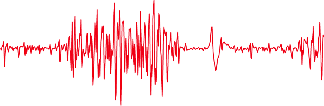
Frequency power spectrum based sEMG signal analysis used by Mpower enables the detection of useful information

An abundance of beneficial information can be identified from sEMG signal with appropriate frequency analysis

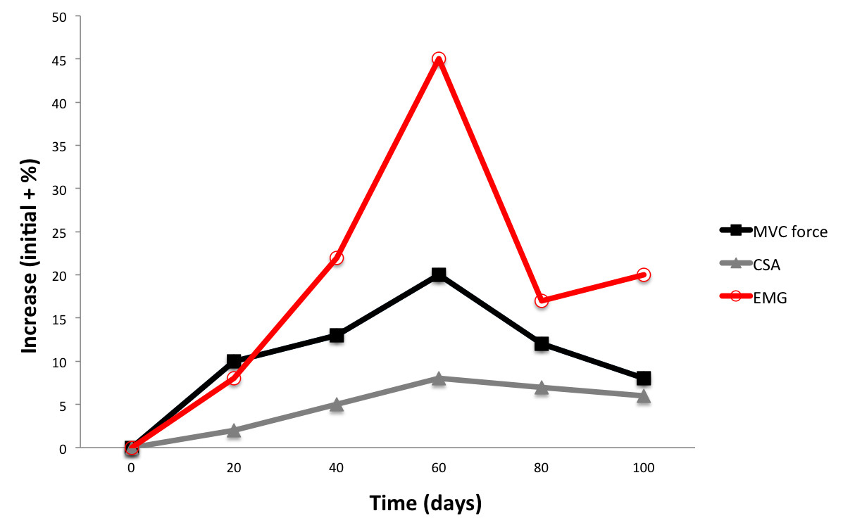
1. Muscle cell type composition and cell size
2. Increasing fast muscle cell activation with faster movements
3. Increasing activation rate of the muscle cells when producing more power
4. Fatigue as a shift of frequency spectrum towards lower frequencies
5. Metabolic changes within the muscle visible in the spectrum

Look for the references >

Referencesclose

o Brody, L. R., et al. (1991). "pH-induced effects on median frequency and conduction velocity of the myoelectric signal." J Appl Physiol (1985) 71(5): 1878-1885.
o Kupa, E. J., et al. (1995). "Effects of Muscle-Fiber Type and Size on Emg Median Frequency and Conduction-Velocity." Journal of Applied Physiology 79(1): 23-32.
o O'Brien, P. R. and J. R. Potvin (1997). "Fatigue-related EMG responses of trunk muscles to a prolonged, isometric twist exertion." Clin Biomech (Bristol, Avon) 12(5): 306-313.
o Potvin, J. R. and L. R. Bent (1997). "A validation of techniques using surface EMG signals from dynamic contractions to quantify muscle fatigue during repetitive tasks." J Electromyogr Kinesiol 7(2): 131-139.
o Wakeling, J. M., et al. (2006). "Muscle fibre recruitment can respond to the mechanics of the muscle contraction." Journal of the Royal Society Interface 3(9): 533-544.
o Contessa, P., et al. (2009). "Motor unit control and force fluctuation during fatigue." J Appl Physiol (1985) 107(1): 235-243.
o De Luca, C. J. and E. C. Hostage (2010). "Relationship Between Firing Rate and Recruitment Threshold of Motoneurons in Voluntary Isometric Contractions." Journal of Neurophysiology 104(2): 1034-1046.
o Gonzalez-Izal, M., et al. (2010). "EMG spectral indices and muscle power fatigue during dynamic contractions." J Electromyogr Kinesiol 20(2): 233-240.
o De Luca, C. J. and P. Contessa (2012). "Hierarchical control of motor units in voluntary contractions." Journal of Neurophysiology 107(1): 178-195.
o Contessa, P. and C. J. De Luca (2013). "Neural control of muscle force: indications from a simulation model." Journal of Neurophysiology 109(6): 1548-

sEMG-signal amplitude measurement results achieved with Mpower correlate extremely well with the corresponding measurements performed with laboratory level measurement equipment. Attached you can find a research report executed by an independent research institute

PDF

Load validation report
/ Chydenius from here

PDF

Load validation report
/ Chydenius from here

Referencesclose